Prioritize Your Heart Health: Understanding Coronary Heart Disease and the Power of Early Detection
February is National Heart Month—a time dedicated to raising awareness about heart health and empowering…
Medical imaging is a crucial tool in modern healthcare, providing detailed visuals of the human body’s internal structures and helping in the accurate diagnosis and treatment of various conditions. At Diagnostic Imaging Northwest (DINW) outpatient centers, patients have access to a variety of imaging modalities, each with specific applications and benefits. In this blog post, we’ll explore the differences and uses of MRI, CT scans, X-rays, ultrasounds and mammography to help you better understand what to expect and how these technologies can assist in your healthcare.
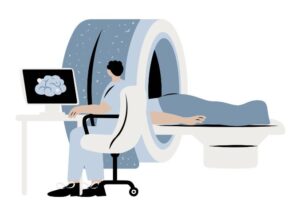 MRI stands for Magnetic Resonance Imaging. Unlike X-rays and CT scans, MRIs do not use ionizing radiation. Instead, they employ a powerful magnetic field along with radio waves to produce detailed images of organs and tissues. This technique is particularly useful for imaging the brain, spinal cord, nerves, muscles, and tendons. Check out the different types of MRI machines we use!
Uses of MRI:
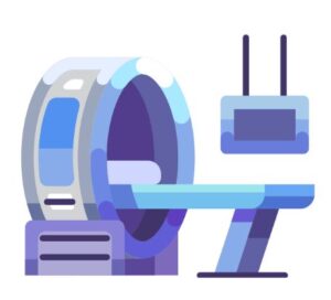 CT scans, or Computed Tomography scans, use X-ray equipment to create detailed images of the body. It combines several X-ray images taken from different angles and uses computer processing to create cross-sectional images of the bones, blood vessels, and soft tissues inside your body. CT scans are faster than MRIs and can be crucial in emergencies.
Uses of CT Scans:
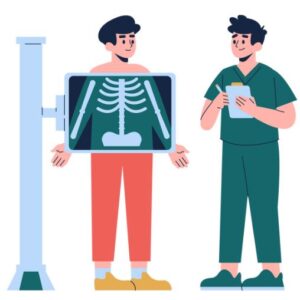 X-rays are a type of radiation known as electromagnetic waves. X-ray imaging creates pictures of the inside of your body — the images show different parts in varying shades of black and white. This is because different tissues absorb different amounts of radiation. X-rays are fast, painless, and commonly used.
Uses of X-rays:
 Ultrasound imaging, also known as sonography, uses high-frequency sound waves to produce images of structures within your body. The images can provide valuable information for diagnosing and treating a variety of diseases and conditions.
Uses of Ultrasound:
 Mammography is a specialized medical imaging technique used to examine breast tissue for signs of abnormalities, including breast cancer. At DINW, we offer 3D mammography, or digital breast tomosynthesis, which provides a more detailed and clearer view compared to traditional 2D mammography.
How 3D Mammography Works:
3D mammography takes multiple X-ray pictures of each breast from different angles and uses computer software to reconstruct these images into a series of thin slices. This process allows radiologists to view the breast tissue layer by layer, like pages in a book, significantly reducing the impact of overlapping breast tissue, which can sometimes obscure or mimic malignancies in 2D images.
Uses of 3D Mammography:
Each imaging technique offered at DINW has its unique strengths and is chosen based on the specific medical situation and the part of the body being examined. Understanding these different imaging techniques can help alleviate any apprehension you might have about undergoing them and allow you to appreciate how they contribute to your overall healthcare management. If you have questions about a particular imaging test, it’s always best to discuss with your healthcare provider who can provide guidance tailored to your personal health needs.
Learn more about Our Services offered at Diagnostic Imaging Northwest.